Hillside Dental
For dentists like Dr. Stephen Welna, X-rays are an invaluable diagnostic tool.

When trying to ensure good oral health, saying “aah” in the dentist’s chair is certainly helpful. Dental professionals like Dr. Stephen Welna of Eau Claire’s Hillside Dental can learn a lot from peeking in their patients’ mouths. But they can’t learn everything, and that’s where X-rays come in.
For more than a century, dentists have been using X-rays in their practices, and these days at Hillside Dental the process is entirely digital. This produces images more quickly and easily and with less radiation than old-fashioned film X-rays, Dr. Welna said.
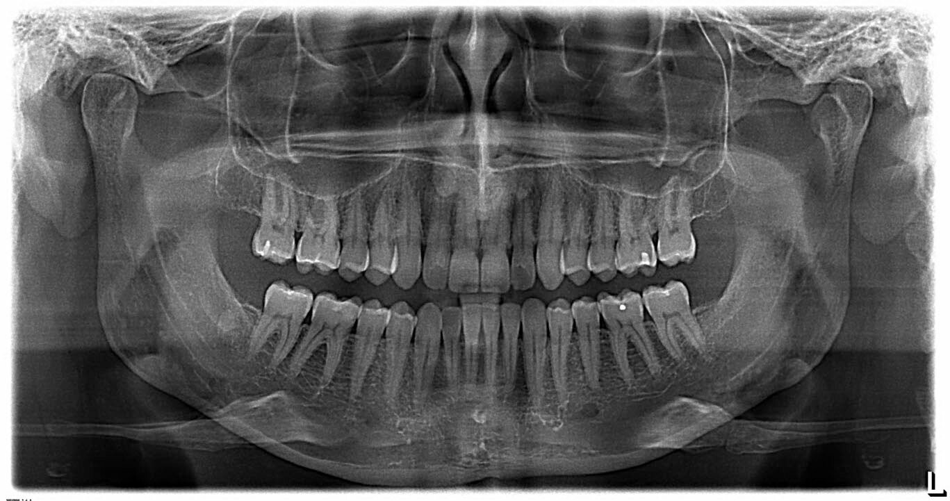
There are three primary kinds of dental X-rays: bitewings, which are normally taken in sets of four and show the back teeth; PA (or periapical) X-rays, which show specific areas of concern, such as a single tooth and its root; and panoramic X-rays, which encompass the whole mouth (see image below of Dr. Welna’s own X-ray). Bitewings are usually taken every year or two, while PA X-rays are taken as needed and panoramic X-rays are taken every five years or so, Dr. Welna said.
X-rays can be used to find out-of-sight cavities, but they’re also useful for much more.
“We always get a lot of questions about radiographs – ‘Are they really necessary? Do we really need to do those?’ – a lot of those kind of questions,” Dr. Welna said. “The quick answer is basically yes: Part of a dental exam is a periodic X-ray examination. We’re looking at the bone, looking at the teeth to make sure there’s no cavities. We’re keeping an eye out for lots of other odd and obscure things that can happen. We’re making sure there’s nothing that looks like it could be potentially cancer. Those kinds of things are at the top of our mind when we’re looking at X-rays.”
Dr. Welna said X-rays can be used to detect bone loss, to identify hard tartar deposits below the gumline, and even in the midst of procedures to make sure they are going as planned.
Yet concerns identified on dental X-rays may not even have anything to do with teeth. Dr. Welna periodically refers patients to their primary-care physicians because he suspects they have blockages in their carotid arteries, which run through the face and neck. And sometimes what a patients thinks is a toothache is actually a problem with their sinuses.
Despite their many benefits, patients can be reluctant to undergo X-rays because they’re worried about the radiation exposure. While he understands such concerns, Dr. Welna said, “At the end of the day, the amount of radiation that we use for dental radiographs is extremely small.”
How small? According to the American Dental Association, the amount of radiation you’re exposed to when you get four bite wing X-rays is about 0.02 millisieverts. In layperson’s terms, that’s less than 1/150ths of the average background radiation exposure an American encounters annually. It’s about one-fifth the amount of radiation in a chest X-ray, 21 times less than the amount in a mammogram, and roughly the equivalent of eating 200 bananas. (Yes, a minuscule amount of the potassium that occurs naturally in bananas is radioactive.)
In other words, it’s only a tiny amount of radiation in exchange for a diagnostic tool that can improve your health — dental and otherwise.
“I always tell patients, half of me putting the pieces of the puzzle together is me looking at the teeth and the other half is the X-ray,” he said. “If you take away half of my tools to do my job, that makes it very challenging to really do things effectively for them.”
Ultimately, one of the best ways to reduce how often you need dental X-rays – and many other dental procedures – is pretty low-tech: brushing and flossing regularly. Patients who take good care of their teeth will need X-rays less often, while those who practice poor oral hygiene might need them more frequently.
“We always tailor what we’re doing with X-rays to the patient,” Dr. Welna said.
![]() Hillside Dental
Hillside Dental
![]() 507 Main St., Eau Claire
507 Main St., Eau Claire
![]() (715) 834-6603
(715) 834-6603
![]() mail@hillsidedental.com(mail (at) hillsidedental [d0t] com,)
mail@hillsidedental.com(mail (at) hillsidedental [d0t] com,)